A 2-year-old girl was resuscitated at Washington Regional Medical Center, Fayetteville, AR, USA, from a Glasgow Coma Scale (GCS) of 3, fixed dilated pupils, and body temperature of 85.1°F (28.9°C) after a 15-minute submersion in 41°F (5°C) water. After 100 minutes of cardiopulmonary resuscitation, the arterial pH was 6.53. Following hypothermia, vasopressors, ventilator support (10 days), and critical care at Arkansas Children’s Hospital, Little Rock, AR, USA, the patient was discharged home 35 days post drowning unresponsive to all stimuli, immobile with legs drawn to the chest, and with constant squirming and head shaking. MRIs at 3 [Figure 1] and 31 [Figure 2] days post drowning showed thalamic injury then generalized atrophy with evolving gray and white matter injury.
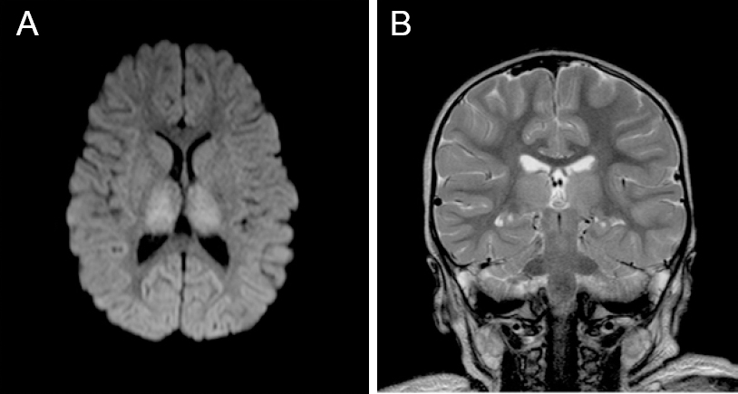
Figure 1
Magnetic resonance imaging at 3 days after injury of the 2-year-old girl who experienced cardiac arrest after cold water drowning.
Note: (A) Axial diffusion-weighted image at three days post drowning, showing increased signal from acute ischemic injury to both thalami; (B) Coronal T2 weighted mid thalamic image 3 days post-drowning, showing subtle diffuse thalamic signal changes, normal ventricles, and normal cortical sulcal cerebrospinal fluid spaces.
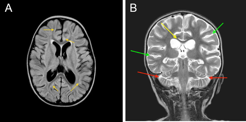
Figure 2
Magnetic resonance imaging at 31 days after injury of the 2-year-old girl who experienced cardiac arrest after cold water drowning.
Note: (A) Axial fluid-attenuated inversion recovery (FLAIR) image at the level of the basal ganglia at 31 days post-drowning, showing subtle persistent diffuse signal irregularities in the gray and white matter (yellow arrows), diffuse gray matter atrophy (enlarged sulcal spaces), and white matter atrophy (enlarged lateral and third ventricles); (B) Coronal T2 image at the level of the thalami 31 days post-drowning, showing gray matter atrophy with increased cerebrospinal fluid spaces at temporal and parietal lobes (green arrows) and cerebellar lobes (red arrows), and white matter atrophy with thinned corpus callosum (yellow arrow) and enlarged ventricles.
Patient had no speech, gait, or responsiveness to commands on day 48 at hospital discharge. She received normobaric 100% oxygen treatment (2 L/minute for 45 minutes by nasal cannula, twice/day) since day 56 and then hyperbaric oxygen treatment (HBOT) at 1.3 atmosphere absolute (131.7 kPa) air/45 minutes, 5 days/week for 40 sessions since day 79; visually apparent and/or physical examination-documented neurological improvement occurred upon initiating each therapy. After HBOT, the patient had normal speech and cognition, assisted gait, residual fine motor and temperament deficits. MRI at 5 months after injury and 27 days after HBOT showed near-normalization of ventricles and reversal of atrophy. Subacute normobaric oxygen and HBOT were able to restore drowning-induced cortical gray matter and white matter loss, as documented by sequential MRI, and simultaneous neurological function, as documented by video and physical examinations.
